How does the normal heart work?
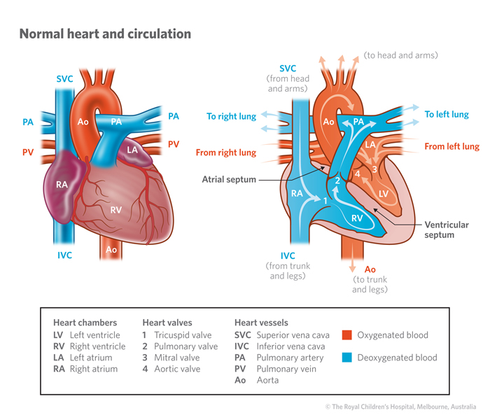
The normal heart is composed of four chambers. The two upper
chambers are reservoirs, which collect blood as it flows back to
the heart. These are called "atriums". From the Atriums blood flows into the
lower two chambers, called "ventricles', which pump blood, with each heart
beat, into the main arteries. From the right side of the heart one
of these arteries (the Pulmonary Artery) carries blood into the
circulation through the lungs. The left side of the heart, on the
other hand, pumps blood into the other main artery (called the
Aorta), which takes blood to the rest of the body.
The two ventricles and the two atriums are separated by
partitions (called Septums). The partition between the Atriums is
called the Atrial Septum (AS) and
that separating the two ventricles the Ventricular Septum (VS). Dark (blue) blood returning to
the right atrium from the body and its organs, through the two main
veins (called the Superior and Inferior Vena Cavas), is pumped by
the right ventricle to the lungs for replenishment with oxygen. The
blue blood becomes bright red in the lungs when oxygen is replaced.
This red blood returns, through two veins from each lung, to the
left atrium and is pumped by the left ventricle to the body
again.

What are the heart
valves?
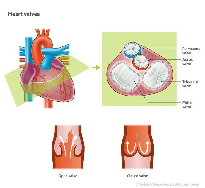
There are four valves which control blood flow through the
heart. They all comprise two or three flaps which swing open to
allow blood through, with each beat, and swing back together to
prevent blood going in the wrong direction. The valves are situated
at the junction of the Atriums with the Ventricles (Tricuspid and
Mitral valves) and at the origin of the major arteries from the
ventricles (Pulmonary and Aortic valves)
The Tricuspid and Mitral Valves are also referred to as
"Atrio-ventricular Valves" while the Aortic and Pulmonary Valves
are called "Arterial Valves".

The heart in a newborn
baby?
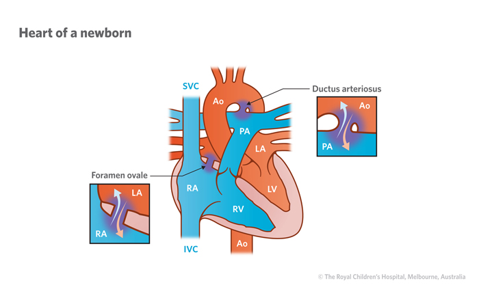
In the newborn infant two communications allow blood to pass
between the two circuits. The "Ductus Arteriosus" is still open
providing a connection between the aorta and the pulmonary artery.
In adition the "Foramen Ovale" allows blood to pass between the
left and right atriums (LA & RA). Within a few days the ductus
closes off completely. The Foramen Ovale closes gradually over
several weeks or months, sometimes remaining open as a tiny slit
into adolescence or beyond.
