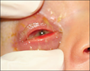
Acute red eye

  • PIC logo
    PIC Endorsed
  • See also

    Eye examination
    Acute eye injury
    Penetrating eye injury
    Periorbital cellulitis

    Key points

    1. Conjunctivitis is the most common cause of acute red eyes
    2. Adequate analgesia will aid examination and procedural sedation may be required in the younger child to allow the clinician to differentiate benign from serious conditions
    3. Key features of sight-threatening conditions are severe eye pain, photophobia, decreased visual acuity or a history of possible penetrating eye trauma; these conditions require specialist ophthalmological assessment

    Background

    • Common causes of red eye include conjunctivitis (viral, bacterial, allergic or chemical), foreign body, corneal ulceration and subconjunctival haemorrhage
    • Uncommon but serious conditions include infectious keratitis, scleritis, uveitis, Penetrating eye injury, endophthalmitis and acute angle closure glaucoma

    Assessment

    History

    • Time course of the redness
    • Eye pain
    • Vision impairment
    • Eye trauma
    • Photophobia
    • Sensation of foreign body (corneal process)
    • Itch
    • Discharge
    • Contact lens wear
    • Fever (see Periorbital cellulitis)
    • Systemic features of inflammation (eg suggestive of Kawasaki disease, PIMS-TS)

    Other factors to consider

    • In a neonate, consider birth history and history of maternal STI
    • Vomiting/coughing
    • Rheumatological/autoimmune conditions or family history
    • Anticoagulant/antiplatelet treatment

    Examination

    See Eye Examination for detailed approach to examination

    Signs and symptoms

    Diagnoses to consider

    Pain, photophobia, watery discharge

    • Foreign body
    • Traumatic corneal ulcer
    • Herpetic ulcer
    • Acute angle closure glaucoma
    • Corneal abrasion

    Purulent discharge

    • Bacterial conjunctivitis
    • Infectious keratitis
    • Endophthalmitis

    Itchiness, eyelid swelling and redness, watery discharge

    • Allergic conjunctivitis
    • Viral conjunctivitis

    Dull, aching eye pain

    • Iritis
    • Scleritis

    Subconjunctival haemorrhage

    • Trauma
    • Vigorous coughing or vomiting

    Focal conjunctival injection

    • Trauma
    • Episcleritis

    Management

    Conjunctivitis
    Infectious, or non-infectious inflammation of the bulbar and/or palpebral conjunctivae

    • Use caution when diagnosing unilateral conjunctivitis

    Bacterial

    Viral

    Allergic

    Presents with purulent discharge, conjunctival inflammation, not pruritic and cornea is clear with no infiltrates

    Treat with eye toilet, topical chloramphenicol 0.5% eye drops 4–6 times per day for 5–7 days. Advise parents of infection control measures (eg washing hands before and after touching eyes)

    If hypersensitivity to chloramphenicol drops stop treatment and consult ophthalmology


    Reproduced with permission from The Royal Australian College of General Practitioners from: Lu SJ, Lee GA, Gole GA. Acute red eye in children: A practical approach. Aust J Gen Pract 2020;49(12):815–22 doi: 10.31128/AJGP-02-20-5240. Available at https://www1.racgp.org.au/ajgp/2020/december/acute-red-eye-in-children 

    Most common type of conjunctivitis

    Presents with watery discharge, burning sensation, bilateral conjunctival inflammation and/or chemosis, and/or eyelid swelling
    Often associated with URTI or gastrointestinal symptoms

    Self-resolving over 1–3 weeks. Symptom control with eye toilet, topical lubricants and cool compresses. Infection control measures. Contagious until eye stops tearing
    A close up of a person's eye  Description automatically generated

    Suspect herpes simplex infection if lid vesicles and/or reduced vision and/or photophobia:

    • Swab for HSV PCR
    • Likely to require treatment with aciclovir
    • Seek specialist advice (ophthalmology, general paediatrics, infectious diseases)
    • If suspected in a neonate, likely to require septic work up. See neonatal conjunctivitis below

    Close-up of a person's eye  Description automatically generated

    Presents with bilateral watery discharge with burning or itchy sensation and eyelid swelling, especially in an atopic child

    Consider treating with antihistamines (oral or topical) and artificial tears

    Specific Conditions

    Condition

    Management

    Neonatal conjunctivitis
    Pathogens include staphylococcus, haemophilus, streptococcus, chlamydia, gonococcus and herpes simplex

    Consider gonococcal conjunctivitis if severe purulent discharge with conjunctival and lid oedema
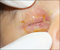
    Figure 1   Figure 2
    Gonococcal ophthalmia neonatorum with swelling, purulent discharge, injection of the conjunctiva and chemosis

    • Obtain conjunctival swabs for gram stain, giemsa stain and cultures, and gonococcal and chlamydia PCR if clinical suspicion
    • Gonococcus, chlamydia and herpes simplex can cause invasive disease and may require septic work up. Seek specialist advice (eg neonatology, infectious diseases, general paediatrics, ophthalmology). See Recognition of the seriously unwell neonate
    • Treat chlamydial conjunctivitis with oral azithromycin 20mg/kg once daily for 3 days and eye toilet
    • Treat gonococcal conjunctivitis with a stat dose of ceftriaxone 25-50mg/kg. Where possible, ceftriaxone should be avoided in neonates <41 weeks gestation, particularly if jaundiced or receiving calcium containing solutions. See Antimicrobial guidelines
    • Herpes simplex infection and/or keratitis is likely to require septic work up and treatment with IV aciclovir. Seek specialist advice
    • Treat other organisms with topical chloramphenicol 0.5% eye drops 4–6 times per day for 5–7 days

    Subconjunctival haemorrhage
    Painless and no discharge
    Associated with activities that increase intraocular pressure; coughing, sneezing, vomiting, valsalva, strangulation

    Potential sign of penetrating eye injuryif history of trauma. If the posterior extent of the haemorrhage cannot be visualised consider an orbital or base of skull fracture

    • In infants, subconjunctival haemorrhage may be a sign of non-accidental injury
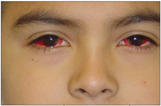
    Case In Point: Subconjunctival Hemorrhages in a Teenage Boy | Consultant360
    Extensive bilateral subconjunctival haemorrhages from excessive vomiting
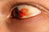

    Subconjunctival haemorrhage of temporal bulbar conjunctiva

    • Observe. If isolated, usually resolves spontaneously within 1 – 3 weeks
    • If recurrent, investigate cause (eg bleeding disorder). Consider non-accidental injury


    Foreign body
    Presents with watery discharge and/or sensation of foreign body
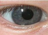
    A close up of a person's eye  Description automatically generated
    Reproduced with permission from The Royal Australian College of General Practitioners from: Lu SJ, Lee GA, Gole GA. Acute red eye in children: A practical approach. Aust J Gen Pract 2020;49(12):815–22 doi: 10.31128/AJGP-02-20-5240. Available at https://www1.racgp.org.au/ajgp/2020/december/acute-red-eye-in-children 

    See Acute eye injury

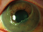
    Corneal abrasion
    Presents with watery discharge and pain that is normally severe initially and then subsides over 24 – 48 hours
    A close up of a person's eye  Description automatically generated with medium confidence A picture containing blue, device, gauge  Description automatically generated
    Reproduced with permission from The Royal Australian College of General Practitioners from: Lu SJ, Lee GA, Gole GA. Acute red eye in children: A practical approach. Aust J Gen Pract 2020;49(12):815–22 doi: 10.31128/AJGP-02-20-5240. Available at https://www1.racgp.org.au/ajgp/2020/december/acute-red-eye-in-children

    See Acute eye injury

    Chemical/thermal burns
    Presents with pain, watery discharge, conjunctival inflammation or pallor in more severe burns and/or corneal epithelial defect and/or corneal opacity
    Reproduced with permission from The Royal Australian College of General Practitioners from: Lu SJ, Lee GA, Gole GA. Acute red eye in children: A practical approach. Aust J Gen Pract 2020;49(12):815–22 doi: 10.31128/AJGP-02-20-5240. Available at https://www1.racgp.org.au/ajgp/2020/december/acute-red-eye-in-children

    • See Acute eye injury
    • Requires urgent, copious irrigation, after local anaesthetic. Sedation or urgent GA may be required


    Infectious keratitis
    Presents with purulent discharge, severe pain, vision loss, intense inflammation of the conjunctiva and corneal opacity (round white spot), or ulcer (dendritic in herpes and varicella) visible after fluorescein staining

    Can be caused by bacteria, viruses, or fungi
    Often associated with contact lens wear
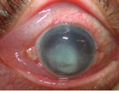
    Reproduced with permission from The Royal Australian College of General Practitioners from: Lu SJ, Lee GA, Gole GA. Acute red eye in children: A practical approach. Aust J Gen Pract 2020;49(12):815–22 doi: 10.31128/AJGP-02-20-5240. Available at https://www1.racgp.org.au/ajgp/2020/december/acute-red-eye-in-children

    • Contact ophthalmology urgently
    • Keep contact lens if possible for culture


    Endophthalmitis
    Presents with severe pain, purulent discharge, vision loss, intense inflammation of conjunctiva and hypopyon

    Risk factors: recent eye surgery, penetrating eye injury, keratitis
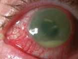
    Reproduced with permission from The Royal Australian College of General Practitioners from: Lu SJ, Lee GA, Gole GA. Acute red eye in children: A practical approach. Aust J Gen Pract 2020;49(12):815–22 doi: 10.31128/AJGP-02-20-5240. Available at https://www1.racgp.org.au/ajgp/2020/december/acute-red-eye-in-children

    • Contact ophthalmology urgently
    • Likely to require broad spectrum IV antibiotics:

    ceftazidime 50mg/kg (maximum 2g) 8 hourly
    and
    vancomycin 15mg/kg (maximum 750mg) 6 hourly

    Episcleritis and scleritis
    Episcleritis presents with eye irritation/mild ocular tenderness, sectoral congestion of episcleral vessels and lacrimation

    Scleritis presents with watery discharge, severe pain, vision loss and intense inflammation of sclera, episcleral and conjunctiva

    Both associated with systemic inflammatory disease

    • Contact ophthalmology (urgent for scleritis)
    • Consider systemic inflammatory disease/autoimmune disease


    Iritis/Uveitis
    Inflammation of the iris (iritis), ciliary body (cyclitis), or choroid (choroiditis)
    Presents with pain, photophobia, blepharospasm and lacrimation

    Pupil may be small, irregular and poorly reactive

    Causes are autoimmune, infection, and trauma

    Contact ophthalmology

    Acute angle closure glaucoma
    Presents with severe eye pain, watery discharge, decreased visual acuity (cloudy vision), cloudy cornea and fixed mildly dilated pupil

    May also have associated headache and vomiting (from raised pressure)

    Contact ophthalmology urgently

    Consider consultation with local paediatric team when

    Any child with suspected child abuse, suspected STI-related conjunctivitis, or possible systemic illness

    Consider consultation with ophthalmology when:

    • A child with a condition present in the table above, or concerns for serious sight-threatening condition
    • If required to facilitate a complete examination particularly in distressed child 

    Consider transfer when

    • A child with a sight threatening condition and/or management is beyond the capacity of the local team 
    • To facilitate ophthalmological assessment

    For emergency advice and paediatric or neonatal ICU transfers, see Retrieval Services

    Consider discharge when

    • Sight-threatening conditions have been excluded AND
    • Appropriate treatment has been initiated as per diagnosis or referral made AND
    • Planned follow up, or clear instructions regarding when early review is indicated has been provided to the child and family

    Parent information

    Conjunctivitis Kids Health fact sheet

    Last updated October 2022

  • Reference List

    1. Gilani, C. Differentiating urgent and emergent causes of acute red eye for the emergency physician. Western Journal of Emergency Medicine. 2017. 18(3), p509-517.
    2. Jacobs, D. Overview of the red eye. UpToDate (viewed 20 April 2022).
    3. Lu, S et al. Acute red eye in children: A practical approach. Australian Journal of General Practice. 2020. 49(12), p815–22. doi: 10.31128/AJGP-02-20-5240. Retrieved from https://www1.racgp.org.au/ajgp/2020/december/acute-red-eye-in-children.
    4. Mahmood, A. Diagnosis and management of the acute red eye. Emergency medicine clinics of North America. 2008. 26(1), p35 – 55.
    5. NSW ACI, Ophthalmology Network, Sehu, W et al. Eye Emergency Manual App. https://aci.health.nsw.gov.au/networks/ophthalmology/about/eem (viewed 19 April 2022).