See also
Resuscitation
Intravenous access - Peripheral
Key points
- Intraosseous access is an effective route for fluid resuscitation, drug delivery and some laboratory evaluation
- Intraosseous access can be used in all age groups and has an acceptable safety profile for emergency vascular access
Background
Intraosseous (IO) access is the recommended technique for
- circulatory access in cardiac arrest
- decompensated shock if vascular access is not rapidly achieved, ie attempts at venous access fail or will take longer than 90 seconds to carry out
The exception is the newborn, where rapid access can be achieved via umbilical vein access or intraosseous access
Contraindications
- Fracture proximal to insertion site
- Vascular injury on the same limb
- Osteogenesis imperfecta
- Overlying skin infection
A burn on overlying skin is not a contraindication to intraosseous access. If possible, choose a site without burns, but intraosseous can often be the only available form of access in the child with severe burns
Potential complications
- Failure to enter the bone marrow, with extravasation or subperiosteal infusion
- Through-and-through penetration of the bone
- Osteomyelitis (rare in short term use)
- Physeal plate injury
- Local infection, skin necrosis, pain, compartment syndrome, fat and bone micro-emboli have all been reported but are rare
Equipment
- Alcohol swabs
- Intraosseous drill (eg EZ-IO®) with needle (sizes pictured below) or manual intraosseous 18G needle with trocar at least 1.5 cm in length
- EZ-IO® connection and EZ stabiliser® dressing (included with needle)
- 3-way extension tap
- 5 mL or 10 mL syringe for aspiration
- 20 mL syringe or infusion pump
- Infusion fluid
- 1% lignocaine if patient is conscious
.png)
Procedure
Identify the appropriate site
 |

|
 |
|
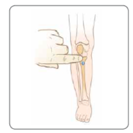
Proximal tibia: Anteromedial surface, 1 finger breadth (~1 cm) below the tibial tuberosity (neonate/young child) or 2 breadths (~2 cm) below the tibial tuberosity (older child) and slightly medial on the flat aspect of the tibia
|
Distal femur: Secure the leg extended 1 cm superior to upper patella border, 1-2 cm medial to midline
|
Distal tibia: 1-2 cm Proximal to the medial malleolus
|
Humerus: Place child’s hand on abdomen with elbow flexed and shoulder internally rotated.� ~1 cm above the surgical neck, on the most prominent aspect of the greater tubercle�Use in children when landmarks can be readily identified
(usually >7 years)
Prepare the skin
Drill insertion
- insert the needle through the skin, perpendicular to the bone, away from the physeal plate. Do not activate drill yet
- at least one black line must be visible outside the skin, confirm adequate needle and set length prior to drilling
- when the needle tip hits bone, press the trigger to commence drilling, applying the minimal amount of pressure required to keep driver advancing into bone
- there is a ‘give’ felt as the marrow cavity is entered. Immediately release trigger
Manual insertion
- insert the needle through the skin perpendicular to the bone, away from the physeal plate
- when the needle tip hits bone, use firm pressure and screwing motion to insert into bone
- there is a ‘give’ as the marrow cavity is entered
Remove the trocar and confirm position by aspirating bone marrow through a 5 mL syringe
- Marrow cannot always be aspirated but intraosseous should flush easily
Secure the needle and start the infusion using 20 or 50 mL syringe manually, or via infusion pump
Laboratory tests
Most laboratory tests cannot be performed on aspirated bone marrow as the particulate matter may block and damage laboratory equipment. Check local laboratory guidelines
Aspirated bone marrow is usually suitable for
- blood cultures
- bedside BSL
- blood gas (in laboratory and some handheld i-STAT instruments)
Ensure to label as marrow specimen
Post-procedure care
Intraosseous infusion should be limited to emergency resuscitation of the child and discontinued as other venous access has been obtained
Perform neurovascular observations on any limb which has had an attempt at intraosseous until
24 hours after removal
Removal
To remove intraosseous needle, remove extension and attach 5 mL syringe to use as handle, pull straight up out of the site, then clean the area
Routine X-ray is not required unless a fracture is suspected
Additional information
Arrow® EZ-IO ® Intraosseous Vascular Access Infant Needle Selection Video
https://www.youtube.com/watch?v=WCoZ3KFSwf0
Consider transfer when
Child requiring care above the level of comfort of the local hospital
For emergency advice and paediatric or neonatal ICU transfers, see Retrieval Services
Last updated December 2024